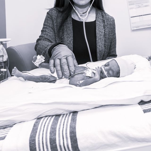
Abdominal Masses in the Neonatal Period
History Gathering
- Oligo or polyhydraminos
- Full-term
- Prenatal US, amniocentesis, pigtail catheter
- Family history
Physical Exam
- Supine : shape, umbilicus, hernias
- Mass: location, size, shape, texture, mobility, tenderness, solid, cystic, air filler
- If distension attempt to relieve to improve exam: NG for obstruction, Crede cath for urinary retention
- +/- rectal exam
- other: chest hypoplasia (Potter’s), bulging hymen (hydrometrocolpos), skin (neuroblastoma)
Differential by location
Flank Mass
-
Renal 55%
-
Hydronephrosis (most common)
- Unilateral: UPJ or UVJ obstruction, aberrant renal artery, kink in ureter
- Bilateral: bladder outlet obstruction (posterior valves or neurogenic bladder) dx: renal scintigraphy and voiding cystourethrogram
- Tx: pyeloplasty, resect obstruction
- Note: sever hydro can be detected in utero and treated with a uretero-amniotic shunt
-
Multicystic Kidney
- Unilateral: multicystic dysplastic kidney
-
Bilateral
- Autosomal recessive polycystic kidney, tx: dialysis, transplant
- Autosomal dominant polycystic kidney presents later
-
Solid kidney tumor
- Most likely mesoblastic nephroma (benign), concentric rings on US, resect
- Wilm’s tumor rarely presents before 6m
-
Hydronephrosis (most common)
-
Juxtarenal
-
Neuroblastoma; most common extracranial malignancy in early childhood, solid, fixed, retroperitoneal (adrenals), catecholamines (sweating, flushing)
- Check urine homovanillic acid, vanillyl mandelic acid
- AXR, US, CT (staging), bone marrow aspiration (look for bone mets)
- Resection +/- chemo and radiation
- Good prognostic factors: less than 1yo, stage, histology
- 4S: stage 1-2 with dissemination to liver, skin (blueberry muffin spots), bone marrow; low risk
- Adrenal hemorrhage – rare, asymptomatic, no management required
- Renal Vein Thrombosis – rare, hematuria, HTN, thrombocytopenia, US, anticoagulation only if bilateral or extending to IVC
- Pulmonary Sequestration
-
Neuroblastoma; most common extracranial malignancy in early childhood, solid, fixed, retroperitoneal (adrenals), catecholamines (sweating, flushing)
Right Upper Quadrant
-
Liver and biliary tree
-
Hemangioendothelioma (aka infantile hepatic hemangioma) - +/- cutaneous hemangiomas, can by asymptomatic or cause AV shunting and CHF, can spontaneously regress
- Part of Kasabach-Merritt syndrome: low platlets, consumptive coagulopathy, hemolytic anemia
- Treat with steroids, IFN, embolization/resection, or transplant
-
Hemangioendothelioma (aka infantile hepatic hemangioma) - +/- cutaneous hemangiomas, can by asymptomatic or cause AV shunting and CHF, can spontaneously regress
-
Hepatoblastoma
- Elevated AFP, associated with Beckwith Wiedermann (macrosomia, macroglossia, visceromegaly, omphalocele)
- Resect, +/- neoadjuvant chemo
-
Hamartoma
- Benign but associated with chromosomal abnl
- Choledochal cyst – cystic dilation of bile ducts, asymptomatic or jaundice, resection and drainage of hepatic duct into intestine
Left Upper Quadrant
- Splenic cyst – resect if large
Mid Abdomen
-
Intestinal
- Pyloric stenois – “olive”, non-bilious emesis, pyloromyotomy
-
Intestinal duplication – cystic or tubular, communicating or non-communicating, often no symptoms but can cause pain, obstruction, bleed, volvulus
- Muscular ring on US, resect
- Intestinal lymphatic malformation (omental, retroperitoneal, mesenteric) – mobile mass, benign but can cause obstruction, resect
-
Meconium
- Ileus – associated with CF, small bowel atresia
- Plug
- Obstruction of prematurity
- Peritonitis or pseudocyst (if perforation occurs)
- Present as distention, doughy mass, ground glass on AXR
- Water soluble contrast enema can be diagnostic and therapeutic
Mid Abdominal Wall
- Fascial defects causing sub-cutaneous mass
- Omphalocele and gastroschisis are not “masses” technically
-
Omphalomesenteric remanents: incomplete vitelline duct reabsorption
- Meckles, omphalomesenteric sinus, cyst, fistula
- Surgical repair
Pelvic
-
Uterine
- hydrocolpos, hydrometrocolpos, imperforate hymen, vaginal transverse septum or atresia (block secretions caused by placental hormones)
Ovarian
- Cystic tumors – mobile, usually benign, if complex on US get markers, large cysts need to be excised or percutaneously drained
- Urachal cyst – the allantois remains and there is a connection between umbilicus and bladder – urine out umbilicus or a cyst
- Teratoma – can cause hemorrhage, polyhydraminos, posterior perineal mass: surgery +/- chemo
-
Hernias and hydrocele
- Inguinal – can incarcerate or strangulate, surgery
- Hydrocele – observe 1 year
Groin mass
- Girls: entrapped ovary
- Boys: ectopic testicle
“The Neonate with an Abdominal Mass” JC Chandler MWL Gauderer, Pediatr Clin Nam 51(2004) 979-997